Fort Lauderdale, Florida – NEW Retail / Consulting Location
2655 East Oakland Park Boulevard, Suite 6, Fort Lauderdale, FL 33306
HOURS: 1-6 PM Monday – Friday ; 1-3 PM Sat. & Sun.
Text 609-489-9425 to schedule appointment for Complimentary Wellness Consulting and / or Purchase of Supplements.
As of 6/4/2024 we are converting ALL our Menu Options at the Top Menu Bar to Clinical Pages, with Clinical Information. Some of the Original Information on those pages will remain at the bottom of the page.
Images will be posted weekly, and below each Image we will discuss briefly a medical / health topic related to the image. If you do not see the brief paragraph following the image, please check back in 7 days. Dr. Miller will post his medical / health topic related to that specific image above.
For those individuals on Immuno-Therapy (Biologics) Infusions for Auto-Immune Disorders, it would be wise to space out your infusions depending on how frequently you are receiving them, in order to prevent extreme suppression f your Immune System.
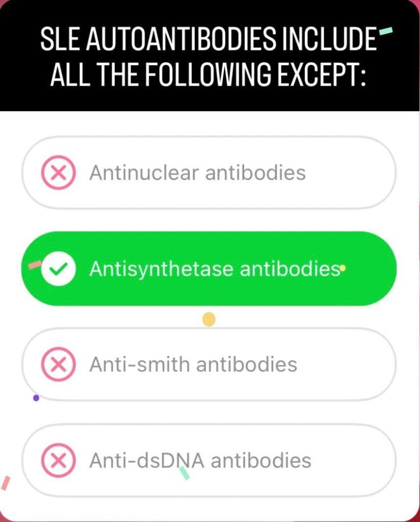
The above question and answers reveals 3 antibodies we test for in people being worked up for possible Lupus (autoimmune disorder). The Antisynthetase antibodies are non-specific antibodies associated with rare auto-immune syndromes and not common in LUPUS. If LUPUS is suspected, antinuclear antibodies, anti-smith antibodies, and anti-dsDNA antibodies should be tested. As with any auto-immune condition, we have immune system dysregulation, with chronic inflammation. It remains critical to lower your inflammation daily, not only to decrease symptoms and flares, however, more importantly to decrease the impact of chronic inflammation on all body tissues and organs from the auto-immune disorder. For Example, with Rheumatoid Arthritis, the joints are not the only tissues affected, as heart muscle, lung tissue, kidneys can all be affected long term by the chronic low level inflammation which persists unrecognized over years, due to the lack of symptoms in those organs initially, and the individual only feeling joint pain.
For those individuals on Immuno-Therapy (Biologics) Infusions for Auto-Immune Disorders, it would be wise to space out your infusions depending on how frequently you are receiving them; in order to prevent extreme suppression of your Immune System. Also, recognize the impotance and therapeutic necessity to lower your inflammation on a daily basis with an anti-inflammatory diet and anti-inflammatory plant-based supplement.
The above image includes: Stomach, Liver, Gallbladder, Pancreas, Small Intestine, Large Intestine and Rectum. The small intestine is Broken up into 3 parts: Duodenum, Jejunum, Ileum. The large intestine is broken up into 4 parts: ascending colon, transverse colon, descending colon, sigmoid colon. The Rectum is the most distal portion of the colon. 2 common autoimmune disorders of the Intestines and Colon are Crohn’s and Ulcerative Colitis. Crohn’s usually involves the last part of the small intestine, Ileum, which connects to the cecum, which is the first part of the large intestine. the apendix comes off the cecum as seen in the image above in the left lower part of the image. However, when referring to the human body and looking at an image, it would be the opposite, that is considered the right lower quadrant of the abdomen. As with any autoimmune disorder, they can affect other distal tissues and organs besides the primary site / organ affected. With Crohn’s we can encounter inflammation of the skin (eczema), eyes, joints (arthritis), iron deficiency anemia, and in children it is important to evaluate for delayed growth, along with other signs and symptoms. Ulcerative Colitis usually involves the sigmoid colon, distal descending colon, and the proximal rectum. The underlying pathophysiology is very similar, inflammation of the wall of the colon. Autoimmune disorders are essentially a dysregulation of the immune system where it starts to react against (attack) a certain subset of tissue. The important aspect to recognize with the intestines and the colon, is we have the advantage of ingesting anti-inflammatory compounds, which will come in direct contact with the lumen of the bowels where most of the pathology exists. Anti-inflammatory plant based compounds are extremely therapeutic for these individuals, especially a high quality curcumin / turmeric supplement. However, there is also pathology in the deeper layers of the wall of the small bowel and colon. As with any organ or tissue, compounds ingested must be absorbed from the small intestines, and transported by the blood to the organ or tissue for proper uptake and utilization. So eventhough we have the direct advantage of the compound coming in direct contact with the pathophysiology in the lumen of the intestines and colon, we need to address the pathology deep in the wall of the intestines and colon, which can only be reached by the same mechanism as with any other organ, absorption of the compounds (such as curcumin / turmeric), and delivery to the bloodstream, then to the organ itself, in this case ALL the layers of the Colon and Intestines. Taking Highly Absorbable and bioavailable plant based vitamins and minerals, rich in anti-oxidants and anti-inflammatory compounds is crucial to control symptoms, flares, and complications of Crohn’s and Ulcerative Colitis.
LOWERING INFLAMMATION (OXIDATIVE STRESS) DAILY, AND AT THE CELLULAR LEVEL, NOT ONLY ELIMINATES EXTERNAL PAIN AND DISCOMFORT, HOWEVER, MORE IMPORTANTLY, IMPROVES THE FUNCTIONING OF ALL YOUR ORGANS AND BODY SYSTEMS.

The main parts of the immune system are:
- white blood cells
- antibodies
- complement system
- lymphatic system
- spleen
- bone marrow
- thymus
White blood cells
White blood cells are the key players in your immune system. They are made in your bone marrow and are part of the lymphatic system.
White blood cells move through blood and tissue throughout your body, looking for foreign invaders (microbes) such as bacteria, viruses, parasites and fungi. When they find them, they launch an immune attack.
White blood cells include lymphocytes (such as B-cells, T-cells and natural killer cells), and many other types of immune cells.
Antibodies
Antibodies help the body to fight microbes or the toxins (poisons) they produce. They do this by recognizing substances called antigens on the surface of the microbe, or in the chemicals they produce, which mark the microbe or toxin as being foreign. The antibodies then mark these antigens for destruction. There are many cells, proteins and chemicals involved in this attack.
Complement system
The complement system is made up of proteins whose actions complement the work done by antibodies. Typically, we measure C3, C4, CH50 levels to help confirm the if someone has immune deficiencies.
Lymphatic system
The lymphatic system is a network of delicate tubes throughout the body. The main roles of the lymphatic system are to:
- manage the fluid levels in the body
- react to bacteria
- deal with cancer cells
- deal with cell products that otherwise would result in disease or disorders
- absorb some of the fats in our diet from the intestine.
The lymphatic system is made up of:
- lymph nodes (also called lymph glands) — which trap microbes
- lymph vessels — tubes that carry lymph, the colorless fluid that bathes your body’s tissues and contains infection-fighting white blood cells
- white blood cells (lymphocytes).
Spleen
The Spleen is a blood-filtering organ that removes microbes and destroys old or damaged red blood cells. It also makes disease-fighting components of the immune system (including antibodies and lymphocytes).
Bone marrow
The Bone Marrow is the spongy tissue found inside your bones. It produces the red blood cells our bodies need to carry oxygen, the white blood cells we use to fight infection, and the platelets we need to help our blood clot.
Thymus
The thymus filters and monitors your blood content. It produces the white blood cells called T-lymphocytes.
Humoral (B-Cell) and Cell-Mediated (T-Cell) Immune Responses
The immune system distinguishes two groups of foreign substances. One group consists of antigens that are freely circulating in the body. These include molecules, viruses, and foreign cells. A second group consists of self-cells that display aberrant MHC proteins. Aberrant MHC proteins can originate from antigens that have been engulfed and broken down (exogenous antigens) or from virus‐infected and tumor cells that are actively synthesizing foreign proteins (endogenous antigens). Depending on the kind of foreign invasion, two different immune responses occur:
The humoral response (or antibody‐mediated response) involves B cells that recognize antigens or pathogens that are circulating in the lymph or blood (“humor” is a medieval term for body fluid). The response follows this chain of events:
- Antigens bind to B cells.
- Interleukins or helper T cells co-stimulate B cells. In most cases, both an antigen and a co-stimulator are required to activate a B cell and initiate B cell proliferation.
- B cells proliferate and produce plasma cells. The plasma cells bear antibodies with the identical antigen specificity as the antigen receptors of the activated B cells. The antibodies are released and circulate through the body, binding to antigens.
- B cells produce memory cells. Memory cells provide future immunity.
The cell‐mediated response involves mostly T cells and responds to any cell that displays aberrant MHC markers, including cells invaded by pathogens, tumor cells, or transplanted cells. The following chain of events describes this immune response:
- Self-cells or APCs displaying foreign antigens bind to T cells.
- Interleukins (secreted by APCs or helper T cells) co-stimulate activation of T cells.
- If MHC‐I and endogenous antigens are displayed on the plasma membrane, T cells proliferate, producing cytotoxic T cells. Cytotoxic T cells destroy cells displaying the antigens.
- If MHC‐II and exogenous antigens are displayed on the plasma membrane, T cells proliferate, producing helper T cells. Helper T cells release interleukins (and other cytokines), which stimulate B cells to produce antibodies that bind to the antigens and stimulate nonspecific agents (NK and macrophages) to destroy the antigens.


